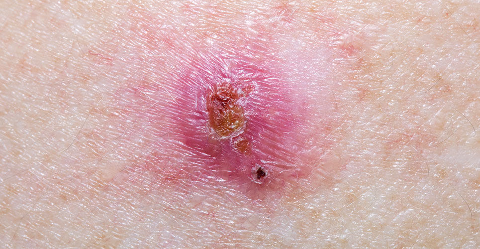
The diagnosis of basal cell carcinoma has been improved over both clinical and dermoscopic examination with a novel imaging technique
Basal cell carcinoma can be diagnosed more accurately than with either clinical or dermoscopic examination using a novel imaging technique according to data presented at the 31st European Academy of Dermatology and Venereology (EADV) Congress by researchers from the Department of Dermatology at the Hôpital Erasme, Université Libre de Bruxelles, Brussels.
Basal cell carcinoma (BCC) is the most frequently diagnosed malignancy worldwide and a Dutch study found that over the period 2001-2019, the age-standardised incidence rates for both men and women with a first BCC increased from 157 to 304 and from 124 to 274 per 100 000 person-years, respectively. Furthermore, the authors of the study concluded that ‘the incidence continues to rise in patients aged 50 years and older. In the next decade a further increase in BCC incidence is expected.’ According to the European Skin Cancer Foundation, in Europe the incidence is of BCC is about 50 to 80 new patients per 100.00 persons per year but which is less than in Australia, where the incidence is about 250 per 100.00 persons per year and with upward trend.
In the current study, researchers found that using a new, non-invasive skin imaging technology called line-field confocal optical coherence tomography (LC-OCT), which gives detailed 3D images at cellular level, significantly increased diagnostic accuracy.
For the differentiation of BCC from BCC-imitators (such as squamous cell carcinoma, actinic and seborrheic keratosis, dermal nevus, and inflammatory conditions), the researchers analysed 303 lesions, including 173 BCC and 130 BCC-imitators. They found that LC-OCT significantly increased the diagnostic accuracy by 12% compared to dermoscopic examination alone (from 85% up to 97%), which the most commonly used skin cancer diagnostic technique.
Importantly, for the differentiation of superficial BCC (a subtype that can be treated non-surgically) from other BCC subtypes, using LC-OCT again increased the diagnostic accuracy by 12% compared to dermoscopic examination alone (from 80% to 92%).
The study also produced a diagnostic algorithm useful to guide the clinician’s diagnosis towards different BCC and BCC-imitators’ subtypes. The algorithm is based on the most powerful LC-OCT morphological criteria that came out from their comprehensive statistical analysis.
Professor Mariano Suppa, a lead researcher and consultant dermatologist from Italy said ‘our findings suggest that, when in front of an BCC equivocal lesion, LC-OCT enables a more accurate diagnosis and, therefore, should be included in the diagnostic process and management of BCC.’ Prof. Suppa added that ‘LC-OCT has the potential to reduce the number of unnecessary biopsies and excisions in cases of superficial BCC and also in the case of benign lesions that do not require surgery.’

