Dark field x-ray enables the identification of structural changes in the lungs of those with COPD that are associated with emphysema.
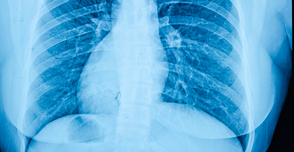
Dark field x-ray enables the diagnosis of emphysema in patients with COPD according to the results of a study by a team from the Department of Physics, Munich School of BioEngineering, Germany. Emphysema is due to the irreversible destruction of alveolar walls, leading to enlargement of distal airspaces and while this leads to changes in the structure of lungs, in the earliest stage it is not possible to detect with a conventional chest x-ray. As x-rays are subject to refraction and ultra small-angle scattering which are not visualised with a conventional X-ray imaging system, in DFX, the contrast produced by these multiple refractions on microstructures in the lungs becomes visible and which enables radiologists to perceive changes that are inaccessible with conventional medical x-ray systems. For example, dark field imaging has permitted the identification of acute lung inflammation in animal models.
With the capacity to determine structural changes at the micro-structural level, the German team undertook their study to examine whether this approach could improve the medical lung assessment in patients with COPD. They included patients with an initial indication of emphysema as revealed by a CT scan but still absent based on spirometry readings. However, a small number of patients with moderate to severe emphysema were also included. COPD classification was based on the post forced expiratory volume in one second (FEV1) and the forced vital capacity (FVC) as proposed by GOLD classification and patients with a FEV1/FVC value of 0.70 were assessed as having no COPD. Dark field x-rays were performed and visually assessed by five readers.
Findings
Overall, 77 patients with a mean age of 65.9 years (male) and 63.3 years (female) were recruited with the majority (83%) at GOLD stage 0. Focusing on the first patient examined, the researchers described how the DFX showed that most parts of the lung yielded dark field values comparable with a healthy lung. A group of 42 patients from the cohort underwent diffusion capacity testing and it was found that the dark field signal gave a strong correlation than with either the CT emphysema index or the visual emphysema grading based on the CT-images.
Commenting on their findings, the authors reported that “in direct comparison, dark-field images and visual evaluation of CT images yield consistent findings regarding emphysema diagnosis.” They also added that the visual features seen with the dark field appeared to provide a greater diagnostic value than conventional emphysema charactering parameters. Moreover, while noting that there are currently no commercially available dark field systems available, their study has shown how the system does not require specialist knowledge and that it is operationally comparable to conventional radiography systems.
In their conclusion, they noted how DRX could offer a low radiation alternative to CT in patients with COPD adding that “x-ray dark-field chest imaging could contribute to improving the detection, diagnosis, and thus treatment and care of pulmonary disorders.”
Citation
Willer K et al. X-ray dark-field chest imaging for detection and quantification of emphysema in patients with chronic obstructive pulmonary disease: a diagnostic accuracy study. Lancet Digit Health 2021