An in vitro study that validates the microbial barrier function of closed infusion systems against microbial touch- and airborne contamination is presented
Healthcare-acquired infections (HAIs) pose a major problem to healthcare systems because they contribute to increased morbidity and mortality in hospitalised patients. Every year, HAIs affect millions of patients worldwide.1,2 In the European Union (EU) alone, the number of HAIs is 4,544,100 per year, leading directly to approximately 37,000 deaths and 16 million extra hospital days.3
Infusion systems are used in daily clinical routine to reconstitute powders and transfer solutions. These infusion systems are expected to be a closed system. However, the meaning of a ‘closed system’ is defined differently in different countries and a common understanding is required.
According to the National Institute for Occupational Safety and Health (NIOSH), a closed system is defined as a device that does not exchange unfiltered air or contaminants with the adjacent environment.4
The International Society of Oncology Pharmacy Practitioners (ISOPP) has separate, independent definitions for microbial closed systems, to prevent microbiological contamination, and chemical closed systems, to prevent the contamination and release of chemicals.5 By taking all different aspects into account, the definitions of a closed system have to be differentiated further:
- Microbial aspect of a closed system:
A device that prevents the transfer of microbial unfiltered air or microbial contaminants into the system - Chemical aspect of a closed system:
A device the prevents the escape of a drug outside the system - Blood aspect of a closed system:
A device that prevents the escape of blood outside the system.
However, in general, the major goal of a closed system is: a) to protect healthcare workers from exposure to infectious agents and potentially harmful drugs; and b) to protect the patient from microbial contamination.
The most common sources of microbial contamination are either a patient’s permanent or transient flora, pathogens from the environment, which is an important aspect in healthcare institutions, antibiotic multi-resistant microorganisms, which have adapted to the hospital environment, or transmission between patients or healthcare staff.6,7
Regarding infection by transmission, the most clinically relevant pathways are direct contact transmission, which can lead to a contamination of infusion systems during handling, and airborne transmission, because the infusion systems are often located for a longer period of time in an unsterile environment, for example, patient rooms, in which airborne transmission cannot be controlled.
Healthcare workers’ hands touch contaminated surfaces during patient care, which increases the risk of direct contact transmission to others.8,9 HAI pathogens that have the potential to spread by the airborne route are also dangerous.10 According to the Centers for Disease Control and the World Health Organization, antibiotic-resistant HAIs are on the rise.
Support for airborne disease transmission is also on the rise.11 Evidence exists for airborne nosocomial transmissions of Acinetobacter, Pseudomonas, and MRSA,12–14 and airborne transmission can spread rapidly and pervasively through a non-immune population.15 Therefore, microbial tightness of infusion systems is an important aspect in preventing microbial ingress and nosocomial infections.
In previous studies, the microbial barrier function has been proven for individual infusion system components used in this study, but only for a single component or connection (unpublished data). Thus, to get closer to clinical reality, the next logical step was the investigation of a complete infusion system as a closed system for microbial contamination with all of its components and handling steps, such as unsealing, disinfection and assembly of the system.
This is the first study investigating the microbial barrier function a complete infusion system as a closed system for microbial hand and airborne contamination.
Aim of the study
The aim of the tests presented here was to prove the microbial barrier function of the connections of a typical infusion system that are exposed to microbial challenge. Two major contamination pathways – touch contamination and airborne contamination – were investigated. The main objective of this study was to evaluate the microbial barrier function of the connection of B. Braun’s infusion system with an Ecoflac® IV container, Intrafix® SafeSet and Caresite® extension set connected to an Introcan Safety® 3 IV catheter.
Microbial barrier test
Two test scenarios were investigated:
- Touch contamination of the connection sites: for this purpose, a defined artificial microbial contamination was used
- Airborne contamination: performed in an aerosol chamber with a defined microbial aerosol.
For testing, a typical infusion system consisting of an Ecoflac® IV container, an Intrafix® SafeSet and a Caresite® Extension set connected to an Introcan Safety® 3 IV catheter was used. The investigations were performed for all components together in a complete assembled infusion system.
Test procedure
All products were tested according to the instructions for use.
- Test setup with microbial contamination of the connection sites
The contamination was carried out with a mixture of three different, but clinical relevant, pathogens. The contamination mixture contained a gram-positive bacteria strain (Staphylococcus epidermidis), a gram-negative bacteria strain (Escherichia coli) and a yeast (Candida albicans).
The number of each species was adjusted to 1 x 10³ colony-forming units (CFU)/ml. For the contamination the connection sites, the ports of the Ecoflac® IV container, the Caresite Valve and the Luer connection of the Introcan Safety® 3 IV cannula, were pressed on a sterile textile moistened with the microbial mixture solution for five seconds. The connection sites were disinfected with a B. Braun Softa® Cloth CHX 2% wipe and air-dried for 15 seconds. The Intrafix® SafeSet was inserted into the Ecoflac® IV container containing 500ml of 0.9ml sodium chloride solution.
A Caresite® extension set was connected to the Intrafix® SafeSet vie the Caresite® valve. Finally, the Caresite® extension set was connected to Introcan Safety® 3 via the Luer connector (Figures 1 and 2). The infusion was administered by gravity flow with a flow rate of approx. 2.5ml/min over a period of approx. 3.5 hours. The infusion solution was collected, centrifuged and analysed for bacterial contamination.
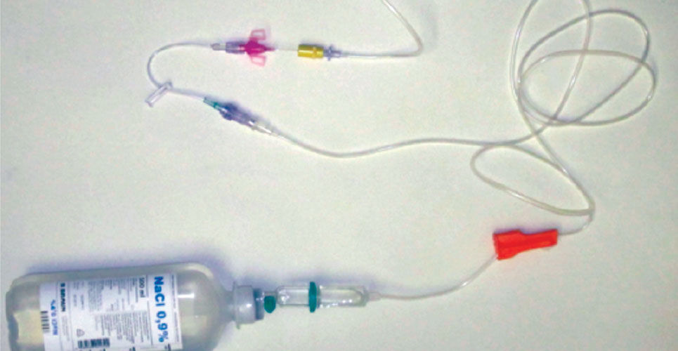
Figure 1. Assembled Infusion setup (outside the sterile clean bench or aerosol chamber for demonstration purposes)

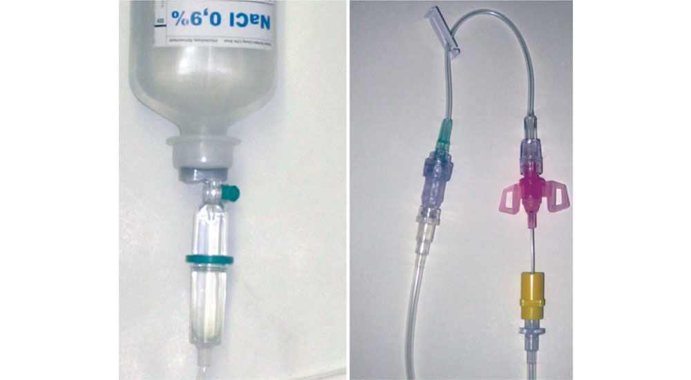
Figure 2. Close-up of assembled connections of Intrafix SafeSet® to Ecoflac® IV container and Intrafix SafeSet® to Caresite® extension line and Introcan Safety® 3 IV cannula (outside the sterile clean bench or aerosol chamber for demonstration purposes)
- Test setup with airborne contamination in an aerosol chamber
The contamination was carried out with a mixture of three different, but clinically relevant, pathogens. The contamination mixture contained a gram-positive bacteria strain (Staphylococcus epidermidis), a gram-negative bacteria strain (Escherichia coli) and a yeast strain (Candida albicans). The number of each species was adjusted to 1 x 10⁵ CFU/ml. All materials were placed inside the aerosol chamber.
A nebuliser containing a suspension of 1 x 10⁵ CFU/ml of each test organism was used to generate an aerosol for 1 minute and 50 seconds. The volume of microbial suspension nebulised per minute was 1ml, which corresponds to an aerosol of 1 x 10⁵ CFU per test organism in the aerosol chamber, which had a volume of 0.25m3. Two small ventilators were used to maintain the aerosol nebula in the chamber over the test period. After five minutes, the Ecoflac® IV container ports were unsealed and the Intrafix® SafeSet, the Caresite® extension set and the Introcan Safety® 3 IV cannula were unpacked. The connection sites were disinfected with a Softa® Cloth CHX 2% wipe and air-dried for 15 seconds. The infusion setup was then connected as described above.
To allow a sterile collection of the infusion solution, the Introcan Safety® 3 was inserted into an IN-Stopper connected to a Heidelberger extension line, which led out of the aerosol chamber into a sterile chamber, in which the infusion solution was collected. The infusion was administered by gravity flow with a flow rate of approx. 2.5 ml/min over a period of approx. 3.5 hrs. The collected infusion solution was centrifuged and analysed for bacterial contamination.
All tests were performed five times with new materials for each single experiment. The average result of all five experiments is reported. An infusion system without disinfection was used as a positive control; a pre-assembled infusion system was used as a negative control.
Results
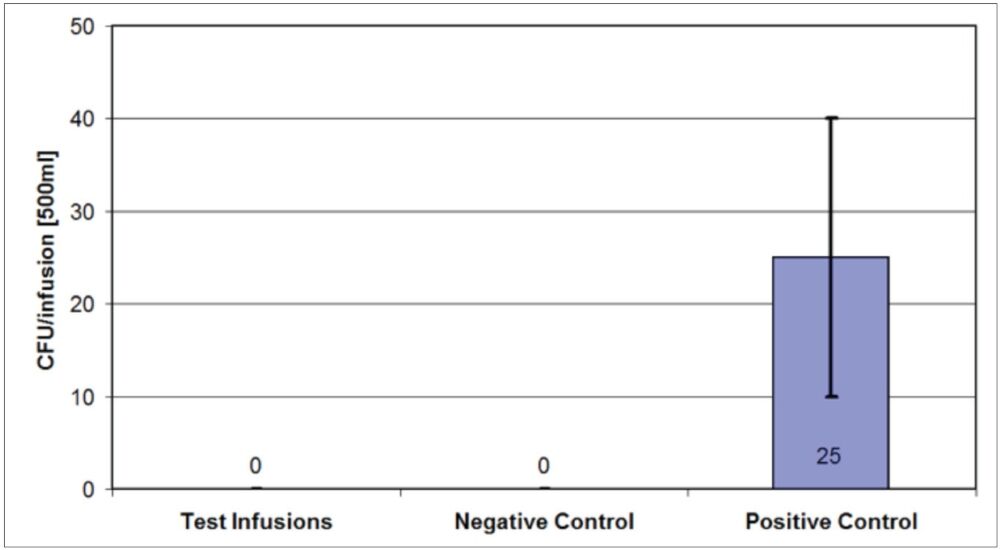
Figure 3 shows that no microbial contamination was detected in all five tested infusion systems after artificial contamination of the connection sites of the infusion system and disinfection prior to assembly of the infusion system. For the positive control, which was contaminated with the microbial solution, but not disinfected prior to assembly, vital organisms have been recovered in the administered infusion solution.

Figure 3. Number of microorganisms isolated from the administered infusion solution after artificial contamination of the connections and subsequent wipe disinfection and assembly of the infusion setup
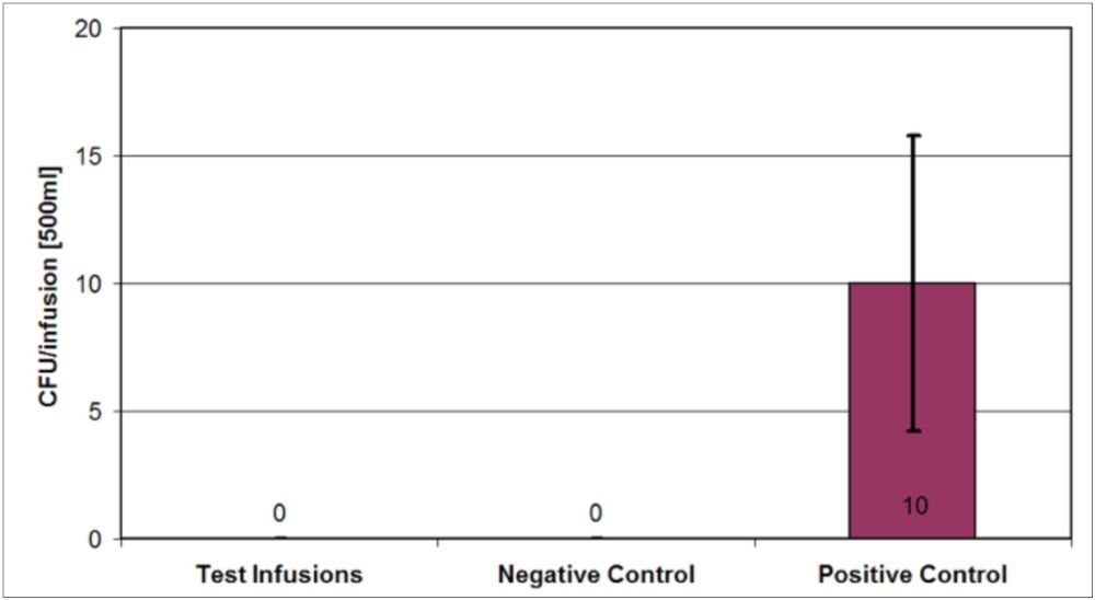
No microbial contamination was detected in all five tested infusion systems after microbial aerosol contamination with 1 x 10⁵ CFU per test strains (Staphylococcus epidermidis, Escherichia coli and Candida albicans; Figure 4). All connection sites were disinfected prior to assembly of the infusion system. A negative control (a pre-assembled infusion system) was used. An infusion system without disinfection prior to assembly was used as a positive control.

Figure 4. Number of microorganisms isolated from the administered infusion solution after aerosol contamination of the connections and subsequent wipe disinfection and assembly of the infusion setup
Discussion and conclusions
The tests were performed in a laboratory setting under controlled conditions. The microorganism mixture comprised one gram-positive strain, one gram-negative strain and one strain of yeast. These groups of microorganisms are normally found in health care settings and HAIs. The data show that the applied test methods are suitable to investigate the microbial tightness of infusion systems. The microbial concentrations of 103 CFU/ml and 105 CFU/0.25m3 are higher than those usually found in hygiene-sensitive areas and, therefore, represent worst-case scenarios. However, the microbial bioburden on fingers, which is one of the most dominant microbial transmission pathways, can vary significantly from person to person.
A relatively high concentration of the microorganisms was used for the aerosol chamber to allow an equal dispersion of organisms in the chamber and a sufficient contamination of the devices used in this study. As seen from the positive controls in both setups, the artificial contamination, the aerosol contamination and the number of recovered microorganisms are relatively low. The reason for this observation is that the presence of microbes on the connection site does not necessarily lead to transmission of the organisms into the infusion system.
Thus, the low number detected in the administered solutions was expected, but also clearly shows that handling without disinfection of the connection sites according to the guidelines can lead to a transmission of microorganisms into the infusion system. Once a transmission occurred, the microorganisms are then infused into the patient’s body and can be the initial step of an infection.
The test methods demonstrate that the tested infusion system components act as a microbial barrier, providing they are applied according to instructions for use and guidelines. The data presented here are the first demonstrating that not only individual parts of an infusion system, but also a complete infusion system, can be classified as a closed system according to the guidelines for hand and airborne microbial contamination.
Declaration of interest
The experiments were carried out on behalf of B. Braun Melsungen. The company did not have any influence on the evaluation of the test results.
Author
QualityLabs BT GmbH, Nuremberg, Germany
References
- Allegranzi B et al. Burden of endemic health-care-associated infection in developing countries: systematic review and meta-analysis. Lancet 2011;377:228–41.
- Bagheri Nejad S et al. Health-care-associated infection in Africa: a systematic review. Bull World Health Org 2011;89:757–65.
- European Centre for Disease Prevention and Control. Annual epidemiological report on communicable diseases in Europe. 2008. www.ecdc.europa.eu/en/publications/publications (accessed December 2016).
- National Institute for Occupational Safety and Health. NIOSH Alert: Preventing occupational exposures to antineoplastic and other hazardous drugs in health care settings. U.S. Department of Health and Human Services, Public Health Service, Centers form Disease Control and Prevention, National Institute for Occupational Safety and Health, DHHS (NIOSH) 2004. Publication No. 2004-165.
- ISOPP. ISOPP Standards of Practice. J Oncol Pharm Pract 2007;13:1–81.
- Weber DJ, Anderson D, Rutala W. The role of the surface environment in healthcare-associated infections. Curr Opin Infect Dis 2013;26(4):338–44.
- World Health Organization. Prevention of hospital-acquired infections. WHO/CDS/CSR/EPH/2002.12; 2002.
- Lemmen SW et al. Distribution of multi-resistant Gram-negative versus Gram-positive bacteria in the hospital inanimate environment. J Hosp Infect 2004;56:191–7.
- Bhalla A et al. Acquisition of nosocomial pathogens on hands after contact with environmental surfaces near hospitalised patients. Infect Control Hosp Epidemiol 2004;25:164–7.
- Kowalski WJ. Aerobiological engineering handbook. McGraw-Hill, New York;2006.
- Fletcher LA et al. The ultraviolet susceptibility of aerosolised microorganisms and the role of photoreactivation. IUVA, Vienna;2003.
- Allen K, Green H. Hospital outbreak of multi-resistant Acinetobacter anitratus: An airborne mode of spread? J Hosp Infect 1987;9:110–19.
- Ryan RM et al. Effect of enhanced ultraviolet germicidal irradiation in the heating ventilation and air conditioning system on ventilator associated pneumonia in a neonatal intensive care unit. J Perinatol 2011;31(9):607–14.
- Farrington M, Ling T, French G. Outbreaks of infection with methicillin-resistant Staphylococcus aureus on neonatal and burns units of a new hospital. Epidem Infect 1990;105:215–28.
- Weinstein RA. Planning for epidemics – The lessons of SARS. N Engl J Med 2004;350(23):2332–4.

