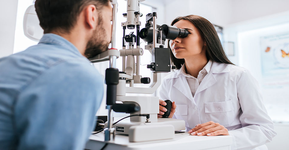
Targeted approaches in the diagnosis and treatment of dry eye were reviewed by an expert international faculty in a satellite symposium at the 3rd EuCornea Congress in Milan, Italy
This content is provided by Allergan Ophthalmology
Dry eye is a prevalent, chronic multifactoral disease of the tears and the ocular surface.(1) Symposium chair, Prof Pasquale Aragona, University of Messina, Italy, highlighted considerations in optimising the tear film and ocular surface system in dry eye syndrome.
Prof Aragona noted that dry eye is accompanied by increased osmolarity of the tear film and inflammation of the ocular surface.(1) Around five million Americans aged 50 years or older have established dry eye, whereas tens of millions more have episodic manifestation due to occupational/environmental tasks or contact lens wear.(2)
An efficient and stable tear film
There are two principal pathogenic pathways of dry eye: aqueous-deficient dry eye, due to insufficient lacrimal gland production and which is recognised as a low-volume hyperosmolar state; and lipid-deficient dry eye, due to alteration of the tear film and lipid layer.(3) Increased evaporation may be seen in both subtypes as disease severity advances.(3)
Prof Aragona explained that tear composition is altered in the course of dysfunctional tear syndrome, marked by decreased concentration of soluble mucins and antibacterial proteins and increased tear osmolarity.(4,5) These changes in tear composition have pro-inflammatory effects on the ocular surface that lead to epithelial damage.(6,7)
Tear hyperosmolarity and tear film instability play key roles in the pathophysiology of dry eye, observed Prof Aragona.(1) “The tear film is central to the ocular surface functional unit, and its efficiency is fundamental to ocular surface homeostasis. Lipids, mucins and aqueous components need to be present and harmonised with each other in order to obtain a stable tear film. The goal of therapy with tear substitutes is to achieve an efficient and stable tear film.”
Aqueous-deficient dry eye
Dr Kostas G Boboridis, Aristotle University of Thessaloniki, Greece, explained that aqueous-deficient dry eye could be thought of as tear-instability dry eye. Tear instability is associated with three pathophysiological events: tear hyperosmolarity; release of inflammatory mediators; and damage to the ocular epithelial cell surface.(1) Epithelial damage involves apoptotic cell death, a loss of goblet cells and disturbance of mucin expression, leading to tear film instability, exacerbation of hyperosmolarity and symptomatic dry eye.(1,8,9)
However, there is often a lack of concordance between signs and symptoms in dry eye.(10,11) This underscores the value of combined diagnostic approaches, incorporating assessment of tear function, lid margin features and the ocular surface. General diagnostic evaluation of dry eye typically entails symptom questionnaires and evaluations of blink rate and blink interval, lower tear meniscus height, tear osmolarity, tear film break-up time, corneal and conjunctival staining and Schirmer test.(1) Osmolarity has been found to have the highest correlation coefficient to disease severity compared with other commonly used diagnostic tests.(12)
Conventional ocular lubricants incorporate a range of viscosity and surface wetting agents designed to support the tear film, for example cellulose derivatives, hyaluronic acid, glycerin containing products, polyethylene glycol/propylene glycol and oil-based emulsion products.(1)
Dr Boboridis outlined extensive evaluations of blended carboxymethylcellulose (CMC) 0.5%, which binds to corneal epithelial cells, is long lasting and supplements the natural mucins of the tear film.(13) In vitro and in vivo evaluations show that CMC accelerated corneal epithelial wound healing in a dose-dependent manner.(13,14)
Dr Boboridis recommended combined treatment approaches that lubricate and also protect the eye through osmoprotection. Elevated tear film osmolarity results in dry eye symptoms, ocular surface damage, upregulation of compensatory mechanisms by epithelial cells and increased apoptosis of epithelial and goblet cells.(15,16) Osmoprotectants such as OptiveTM (Allergan) protect eyes from hyperosmolarity by reducing hypertonicity below the ocular surface, providing hydration to the ocular surface and indirectly controlling the vicious inflammatory cascade.(17)
Conventional hypotonic eye drops lower tear osmolarity but only for a very short time.18 Osmoprotective agents contain compatible solutes (for example, erythritol, L-carnitine, glycerin), which enter corneal cells at a significant rate, protect cells from hyperosmolarity, reduce osmotic stress and thereby influence indirectly inflammatory mediators.(17,19,20)
“Overall treatment goals in aqueous-deficient dry eye are to support the tear film, control osmolarity and reduce inflammation,” Dr Boboridis concluded. “Intervention is designed to preserve the ocular surface and tear film, thereby improve ocular comfort and quality of life of individual patients.”
Lipid-deficient dry eyes
Challenges and solutions in the management of lipid deficient or evaporative dry eye were addressed by Dr Elisabeth Messmer, Ludwig-Maximilians-University, Munich, Germany.
Lipid deficient dry eye is a common condition.(21-23) The key pathogenic mechanism is meibomian gland dysfunction (MGD), a chronic, diffuse abnormality of the meibomian glands leading to reduced or altered meibum at the lid margin and in the tear film.(14,16) Lipid deficiency or alterations may lead to increased evaporation, hyperosmolarity and instability of the tear film, increased bacterial growth on the lid margin, evaporative dry eye and ocular surface inflammation and damage.(22)
Careful clinical history and symptom assessment are vital first steps in the diagnosis of MGD-related dry eye.(22)
A sequence of clinical tests should be carried out to refine diagnosis, classification and grading.(24,25)
Dr Messmer recommended evaluation of eyelid margin (for example, malpositioning, anterior blepharitis, hyperkeratinisation), orifices (for example, number present and number plugged), the quality of expressed meibum and meibum expressibility, meibomian gland mass (for example, using non-contact meibography), tear film stability and measurement of tear osmolarity (if available). Hyperosmolarity of the tear film is an important pathogenetic factor in dry eye.(26) Clinicians were encouraged to check also for signs of associated aqueous-deficient dry eye.
Although MGD is considered likely to be the most common cause of dry eye, it remains underdiagnosed and is often associated with improper follow up when detected, noted Dr Messmer. The major goal in the treatment of MGD is improvement in the patient’s symptoms.
A mainstay of therapy is intensive patient education regarding the chronic nature of the condition and lid hygiene.(25)
“I advise hot compresses for five minutes morning and evening,” said Dr Messmer. “Thereafter, lid massage with closed lids, massage upper and lower lids with a wet cotton tip towards the lid margin.”
Depending on clinical severity, therapy for MGD-related dry eye may involve artificial tears with lipid components, topical antibiotics to the lid margin, topical corticosteroids, systemic tetracycline derivatives and omega-3 fatty acid supplements.(25,27) “A short course of topical antibiotics is often necessary in many MGD patients,” said Dr Messmer. “Systemic tetracycline derivatives (for example, doxycycline 50-100mg once or twice daily) are helpful in reducing inflammation and bacterial load in the lids. Omega-3 fatty acid supplements may also be considered as complementary anti-inflammatory anti-apoptotic therapy.”
Osmoprotectant artificial tears with lipid components designed specifically to restore osmolarity and reduce tear evaporation offer a new and effective topical treatment option for MGD and associated lipid-deficient dry eye, Dr Messmer observed. Optive Plus contains a low concentration of a highly purified lipid derived from the seeds of the castor oil plant, facilitating optimal spreading on the tear film.(28-31)
Surgical techniques, such as meibomian gland intraductal probing, also show promise and warrant further clinical investigation, Dr Messmer concluded.(32)
A dynamic approach works best
The symposium closed with a lively Q&A session involving delegates and faculty. Panel discussants commented that, in the absence of signs or symptoms of dry eye, clinicians should not rely on Schirmer test results alone in diagnosing dry eye.
Inflammation is common to both aqueous-deficient dry eye and lipid-deficiency MGD. Clinicians should aim for maximum control of inflammation to prevent end-stage hyperkeratinisation of the glands, Dr Messmer observed.
“Dry eye is a dynamic disease and treatment approaches should be dynamic too,” remarked Prof Aragona. “Signs and symptoms of dry eye alter over time and in response to therapy. Choice of therapy is made following an exact diagnosis and precise staging of the disease. Treatment of dry eye should address aqueous and lipid deficiency to effectively relieve clinical symptoms.”
References
- DEWS. Ocul Surf 2007;5(2):67-204.
- Dogru M, Tsubota K. Expert Opin Pharmacother 2011;12(3):325-34.
- Bron AJ et al. Ocul Surf 2009;7(2):78-92.
- Solomon A et al. Invest Ophthalmol Vis Scii 2001;42(10):2283-92.
- Ogasawara K et al. Graefes Arch Clin Exp Ophthalmol 1996;234(9):542-6.
- Stern ME, Pflugfelder SC. Ocul Surf 2004;2:124-30.
- Stern ME et al. Exp Eye Res 2004;78(3):409-16.
- Bron AJ et al. Ocul Surf 2011;9(2):70-91.
- Knop N et al. Cornea 2012;31(6):668-79.
- Nichols KK, Nichols JJ, Mitchell GL. Cornea 2004;23(8):762-70.
- Lemp MA. Am J Ophthalmol 2008;146:350-6.
- Sullivan BD et al. Invest Ophthalmol Vis Sci 2010;51:6125-30.
- Garrett Q et al. Invest Ophthalmol Vis Sci 2007;48(4):1559-67.
- Garrett Q et al. Curr Eye Res 2008;33:567-73.
- Yeh S et al. Invest Ophthalmol Vis Sci 2003;44(1):124-9.
- Luo L, Li DQ, Pflugfelder SC. Cornea 2007;26(4):452-60.
- Corrales RM et al. Cornea 2008;27(5):574-9.
- Holly FJ, Lamberts DW. Invest Ophthalmol Vis Sci 1981;20(2):236-45.
- Xu S et al. Mol Vis 2010 Sep 4;16:1823-31.
- Garrett Q et al. Invest Ophthalmol Vis Sci 2008;49(11):4844-9.
- Lemp MA et al. Cornea 2012;31(5):472-8.
- Nichols KK et al. Invest Ophthalmol Vis Sci 2011;52:1922-9.
- Schaumberg DA et al. Invest Ophthalmol Vis Sci 2011; 52(4):1994-2005.
- Foulks GN et al. Ophthalmology 2012;119(10 Suppl):S1-12.
- Geerling G et al. Invest Ophthalmol Vis Sci 2011;52(4):2050-64.
- Messmer EM, Bulgen M, Kampik A. Dev Ophthalmol 2010;45:129-38.
- Macsai MS. Trans Am Ophthalmol Soc 2008;106:336-56.
- Di Pascuale MA, Goto E, Tseng SC. Ophthalmology 2004;111(4):783-91.
- Maïssa C et al. Cont Lens Anterior Eye 2010;33(2):76-82.
- Khanal S et al. Cornea 2007;26(2):175-81.
- Kaercher T, Buchholz P, Kimmich F. Clin Ophthalmol 2009;3:33-9.
- Maskin SL. Cornea 2010;29(10):1145-52.

