teaser
Christoph Abels
MD PhD
Clinical Associate Professor
Department of Dermatology
University of Regensburg
Medical Director
August Wolff GmbH & Co KG Arzneimittel
Bielefeld
Germany
E:[email protected]
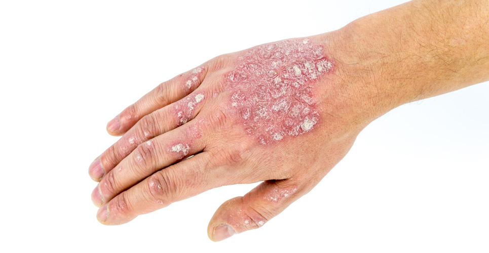
Atopic dermatitis or atopic eczema is defined as a chronic or chronic relapsing, noncontagious, eczematous skin disease accompanied by severe itching.(1) The localisation of the disease changes during the patient’s lifetime; however, in infants and adults the flexures are typically affected (see Figure 1). Clinical symptoms are erythema with papulovesicular lesions with crusting in acute stages, whereas in chronic stages pruriginous lichenoid papules and lichenification prevail.(2)
[[HPE30_fig1_46]]
Recently, the World Allergy Organisation suggested to engulf all local inflammatory reactions of the skin under the term “dermatitis”, which will cover contact dermatitis, eczema and other types of dermatitis. In addition, the term eczema comprises atopic eczema with IgE-levels >150kU/l and nonatopic eczema with IgE-levels <150kU/l and without association with allergic asthma or rhinoconjuctivitis.(3)
The prevalence of atopic eczema has been constantly increasing over the last 50 years. Currently, the prevalence in school beginners is estimated to be 8−16% in western European countries. Among adults the prevalence is approximately 3%. The genetic background of the disease is well established.(1,4,5)
The pathogenesis and pathophysiology of atopic eczema is complex and still not completely clear. One assumes a combination of different disorders, such as:
- Epidermal barrier disorder.
- Humoral and cellular immunity disorder.
- Antigen-presenting cells of the epidermis disorder.
- Vegetative nerve system disorder.
However, and most importantly, the physiological epidermal barrier is disturbed in atopic eczema. For some time the disturbance of the epidermal barrier was seen as a consequence of the inflammatory phenotype (“inside-out” hypothesis). However, several investigations in recent years have pointed towards a genetic disorder affecting epidermal differentiation of the keratinocytes as the underlying cause for barrier disturbance in atopic eczema.(6-9) Therefore, irritant or allergenic substances may enter the living epidermis and induce inflammation and epidermal hyperplasia in atopic individuals (“outside-in” hypothesis).(9)
Increased transepidermal water loss and reduced capacity to bind water in the epidermis are characteristic of a disturbed epidermal barrier. In a recent study it was shown that, in atopic eczema, mutations of the filaggrin gene are present.(10) This can explain the dry skin observed in atopic patients, because products with a filaggrin metabolism are important in the water-binding process of the stratum corneum.
Another important role for the maintenance of the epidermal barrier is the presence of the unsaturated fatty acid linoleic acid. Unsaturated fatty acids such as linoleic acid or γ-linolenic acid are polyun saturated ω-6 fatty acids, which are part of the intercellular lamellar lipid structure of the stratum corneum, which contributes significantly to the barrier function of the epidermis. In addition, polyunsaturated fatty acids are constituents of ceramides 1 and 4. A lack of these essential fatty acids in the daily diet can lead to eczematous skin with disturbed epidermal barrier in both humans and animals.(11,12)
Topical therapy
Due to the disturbed epidermal barrier in atopic eczema, emollients or moisturisers are still the mainstay for the treatment of atopic eczema. Ronald Marks described their role in dermatology in a metaphor: “They are a bit like buttons on a shirt – important for adequate function but don’t do much to keep you warm.”(13)
Lipid-rich emollients may reconstitute the epidermal barrier, therefore reducing hyper proliferation and inducing epidermal differentiation. Consequently, the water-binding capacity of the stratum corneum is increasing and the transepidermal water loss reduced. Finally, the ability of irritant or allergenic substances to enter the skin is reduced and the amount of corticosteroids is cut down.(14,15)
However, there is no cream or ointment for every case. Therefore, the emollient must be adapted to the stage of the atopic eczema, the age of the patient, the localisation and the season (see Table 1).
[[HPE30_table1_47]]
Another important aspect of topical therapy of atopic eczema is the amount of emollient used by the patient. In general, the amount needed is underestimated by patients as well as by physicians. To treat the entire body, 20g per application are needed. In a recently published study it was found that patients only used 2g or less.(16) To educate patients on the amount of emollient to apply, the “fingertip unit” may be used.(17) A fingertip unit for a male adult represents 0.5g, for a female adult 0.4g and for children at the age of four approximately one-third that of an adult.
If topical emollients are insufficient to keep the atopic eczema under control, topical cortico steroids and the calcineurin inhibitors (pimecrolimus and tacrolimus) should be used.(1,18-20) Despite the extensive discussion of the potential side-effects of corticosteroids – in particular in the lay press – these anti-inflammatory drugs are still the first-line treatment. Potential side-effects are minimised if a more powerful corticosteroid is used for as short a time as possible and then a less powerful corticosteroid is chosen as soon as possible. Another possibility to reduce the potential side-effects of cortico steroids and prevent flare-ups is intermittent use, for example, once-daily twice-weekly, as demonstrated in a recent study in combination with emollients.(21) After healing, the patient should switch back to the preferred emollient containing, for example, unsaturated fatty acids or urea. The calcineurin inhibitors pimecrolimus and tacrolimus provide additional weapons to fight atopic eczema, but only as second-line drugs (see Table 2).
[[HPE30_table2_48]]
Conclusion
Despite the introduction of the calcineurin inhibitors, emollients and topical corticosteroids remain the mainstay in the treatment of atopic eczema. In particular, emollients with hardly any side-effects may attenuate the disease and even reduce the number of flare-ups if used consequently and sufficiently. If atopic eczema is not controlled by emollients alone, corticosteroids should be used as early as possible or calcineurin inhibitors as a second-line option. The education of patients suffering from atopic eczema on how and when to use their OTC or prescription drugs is the key to success.
References
- Leitlinien der Deutschen Dermatologischen Gesellschaft (DDG) und des Berufsverbandes Deutscher Dermatologen (BVDD) (2002) Atopische Dermatitis Available from: http://www.awmf-online.de
- Williams HC. Atopic dermatitis.N Engl J Med 2005;352: 2314-24.
- Johansson SG, Bieber T, Dahl R,et al. Revised nomenclature for allergy for global use: report of the �nomenclature review committee of the World Allergy Organisation. J Allergy Clin Immunol 2004;113: 832-6.
- Wei�buch Allergie in Deutschland, 2. Aufl. M�nchen, Germany: Urban & Vogel; 2004.
- Luoma R, Koivikko A, Viander M. Development of asthma, allergic rhinitis and atopic dermatitis by the age of five years: a prospective study of 543 newborns. Allergy 1983;38: 339-46
- Cookson W. The immunogenetics of asthma and eczema: a new focus on the epithelium. Nat Rev Immunol 2004;4:978-88.
- Jensen JM, Folster-Holst R,Baranowsky A, et al. Impaired sphingomyelinase activity and �epidermal differentaiation in �atopic dermatitis. J Invest Dermatol 2004;122:1423-31.
- Proksch E, Jensen JM, Elias PM.Skin lipids and epidermal �differentiation in atopic dermatitis.Clin Dermatol 2003;21:134-44.
- Strid J, Strobel S. Skin barrier dysfunction and systemic sensitization to allergens through the skin. Curr Drug Targets Inflamm Allergy 2005;4: 531-41.
- Palmer CN, Irvine AD, Terron-Kwiatkowski A, et al. Common loss-of-function variants of the �epidermal barrier protein filaggrin area major predisposing factor for atopic �dermatitis. Nat Genet 2006; 38:441-6.
- Horrobin DF. Essential fatty acid metabolism and its modification in atopic eczema. Am J Clin Nutr 2000;71 Suppl 1:367-72.
- Proksch E. Linolsaurehaltige Externa. Theoretische Grundlagen und praktische Bedeutung. Internistische Praxis 1998;38:877-83.
- Marks R. Sophisticated emollients. Stuttgart, Germany: Thieme; 2001.
- Ekanayake-Mudiyanselage S, Aschauer H, Schmook FP, et al. Expression of epidermal keratins and the cornfied envelope protein involucrin is influenced by permeability barrier disruption. J Invest Dermatol 1998;111:517-23.
- Proksch E, Feingold KR,Elias PM. Barrier function regulates epidermal DNA-synthesis.J Clin Invest 1991;87:1668-73.
- Niemeier V, Kupfer J, Schill WB, Gieler U. Atopic dermatitis – topical therapy: do patients apply much too little? J Dermatol Treatment 2005;16: 95-101.
- Long CC, Finlay AY. The finger-tip unit – a new practical measure.Clin Exp Dermatol 1991;16:444-7.
- Gemeinsamer Bundesausschuss. Therapiehinweis nach Nr. 14 der Arzneimittelrichtlinien. Tacrolimus zur topischen Behandlung. Dt. Arzteblatt 2004;101:A529-31.
- Gemeinsamer Bundesausschuss. Therapiehinweis nach Nr. 14 der Arzneimittelrichtlinien. Pimecrolimus zur topischen Behandlung. Dt. Arzteblatt 2004;101: A601-3.
- Hornstein OP, Nurnberg E. Externe Therapie von Hautkrankheiten. Stuttgart, Germany: Thieme; 1985.
- Berth-Jones J, Damstra RJ,Golsch S, et al. Twice weekly fluticasone propionate added to emollient maintenance treatment to reduce risk of relapse in atopic dermatitis: randomised, double blind, parallel group study. BMJ 2003;326: 1367-72.

