teaser
Jann Lübbe
MD
Head of Atopic Dermatitis Outpatient Consultation
Dermatology Clinic
University Hospital
Geneva
Switzerland
E:[email protected]
The advent of new topical immunomodulators such as tacrolimus and pimecrolimus has led to renewed interest in the treatment of atopic dermatitis, as these drugs represent the first valuable alternative to topical corticosteroids, which have been the backbone of atopic dermatitis management for almost 50 years.
The main incentive to develop topical calcineurin inhibitors was to combine the anti-inflammatory effectiveness of their systemic counterparts (such as ciclosporin and tacrolimus) with a reduced side-effect profile, so that they could be used in daily practice. This was achieved with the development of topical preparations of tacrolimus and pimecrolimus, which are now registered in most western countries; numerous clinical trials have demonstrated the efficacy and safety of these preparations in the treatment of atopic dermatitis in children and adults.(1–4) The role of these new treatments in daily practice will depend on long-term experience in clinical practice. Topical calcineurin inhibitors are not a causal cure for atopic dermatitis, and sustained success with any treatment in a chronic relapsing disease such as atopic dermatitis still depends on good know-how in basic skincare. Therefore, the current widespread interest in the new topical calcineurin inhibitors is an excellent occasion to review the essentials of topical treatment in atopic dermatitis. Moreover, the exaggerated expectations and fears that are associated with any groundbreaking new treatment should be addressed with medical common sense: topical corticosteroids will not become obsolete, but will rather be used in combination with topical calcineurin inhibitors in the management of atopic dermatitis. Irrational fears of corticosteroids should not be exploited in order to promote topical calcineurin inhibitors. On the other hand, fears related to the long-term adverse effects of calcineurin inhibitors, especially with regard to UV-induced skin carcinogenesis, should be discussed in an open and rational way.
Principles of topical therapy
In most cases of atopic dermatitis, systemic treatment can be avoided, provided that the patient complies with the time-consuming and inconvenient topical therapy. Educating patients and/or parents is essential, as the principles of topical treatment apply to both flare management with medicated preparations and baseline daily skincare with unmedicated emollients. The appropriate amount of product to be spread on the skin is often underestimated. For example, one course of treatment for the back of an adult patient requires 7–9g of cream, and treatment of the head and neck 3–4g. The fingertip rule (see Figure 1) is a useful way to estimate the appropriate dosage in children and adults.(5) Topical treatment should always be applied on hydrated skin (ie, immediately after a bath or shower), before the skin dries up and becomes tense again. Hydration increases the permeability and absorbance of the stratum corneum and reduces the mechanical stress of spreading, which can increase irritation of an already inflamed skin. This is especially important when using ointments. Patients with acute, exudative and erosive lesions, in particular children, often do not tolerate standard topical application and may be treated first with “wet wraps” until the oozing stops (which usually happens after two or three applications). Wet wraps are made of ointment-coated tissue soaked in physiological saline solution or water.(6) However, there is a risk of secondary viral and bacterial infection, and the possibility of increased systemic absorption and skin atrophy requires close monitoring when medicated creams are used.
Topical therapy is time-consuming, even if wet wraps are not used: patients should plan 30 minutes for each session. One well-conducted treatment per day is usually sufficient in the treatment of active disease, and unmedicated baseline therapy must be a daily habit in disease-free intervals.
[[HPE18_fig1_21]]
Anti-inflammatory treatment
Topical calcineurin inhibitors
The main advantage of calcineurin inhibitors over topical corticosteroids is that they do not induce skin atrophy,(7,8) which makes them the first-line treatment for sensitive skin areas such as face and skin folds, and a promising option for topical long-term management. Tacrolimus is available as a 0.03% and 0.1% ointment, and pimecrolimus as a 1% cream. The choice of either treatment in clinical practice depends on skin tolerance and required anti-inflammatory potency. The potency of tacrolimus 0.1% ointment is similar to that of a group III corticosteroid,(9) which is more than that of 1.0% pimecrolimus cream.(10) The potency of pimecrolimus cream 1.0% is comparable to that of 0.03% tacrolimus ointment,(11) which is clearly more than a group I corticosteroid.(12) However, pimecrolimus cream is much better tolerated and cosmetically more acceptable.(1–4,12) Patients should be informed of the possible occurrence of a transitory local intolerance reaction characterised by intense burning and itch; this reaction is experienced by up to 50% of patients using tacrolimus ointment,(2,3) versus 10% of patients using pimecrolimus.(1,4)
Systemic absorption of the drugs during treatment is minimal and does not constitute a problem in daily practice, but, as a general rule, treatment should be discontinued in case of persistent treatment failure and occlusive treatment should be avoided. Significant persistent blood levels of tacrolimus have been found in the context of sustained skin barrier abnormalities such as the Netherton syndrome.(13)
There is no statistical evidence for an increased risk of skin infections during treatment with topical calcineurin inhibitors, and staphylococcal counts decrease during local therapy.(14) However, extensive viral superinfection with herpes simplex(15) or molluscum contagiosum(16) has been observed, and patients should be instructed to discontinue therapy if unusual lesions develop.
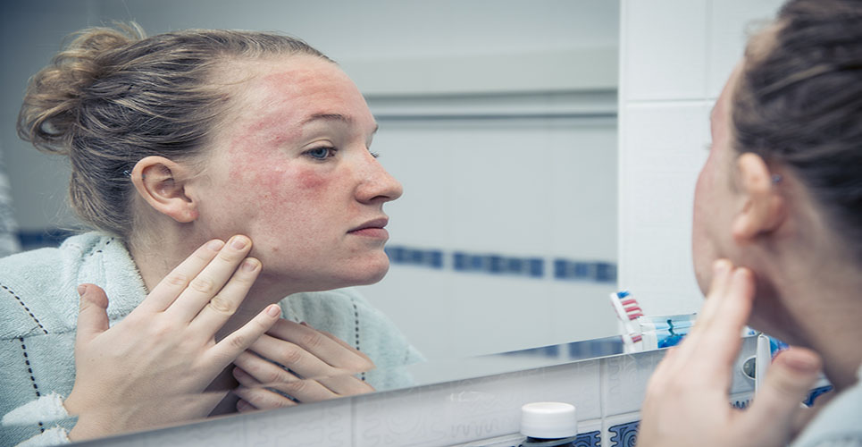
Alcohol intolerance, which is observed more often with tacrolimus, manifests as facial flushing after intake of even minimal quantities.(17,18) Rosaceiform dermatitis has been observed as a rare side-effect of facial treatment with both pimecrolimus and tacrolimus.(19,20)
No study has yet been carried out on concomitant treatment with topical corticosteroids, but randomised studies have assessed the utility of steroids for the management of flares failing to respond to pimecrolimus cream.(1,4)
The drugs have no photosensitising potential, and ordinary photoprotection is sufficient during treatment; concomitant phototherapy, however, should be avoided.
Long-term safety
Studies still need to be carried out to establish the long-term safety profile of topical calcineurin inhibitors. Concerns(21) stem from the well-documented effects of systemic immunosuppression with ciclosporin or tacrolimus on the development of viral papillomas and skin cancer on photoexposed skin.(22) Studies in animal models have shown controversial results.(23) As no representative animal model for human UV-related carcinogenesis is available, conclusions will rely on prospective observations.
There is currently no clinical or experimental evidence for a local carcinogenic effect of topical calcineurin inhibitors, in spite of prospective studies with up to four years of follow-up, and even more in single cases.(24) This fact should be put into perspective with regard to other well-established treatments for atopic dermatitis for which a carcinogenic risk is recognised and accepted, such as phototherapy.
Topical corticosteroids
Topical corticosteroids have been the mainstay of anti- inflammatory treatment for almost half a century, and, due to the resulting predictability of benefits and risks, they remain the first choice for the treatment of exacerbated atopic eczema.(24) The widespread fear of using corticosteroids is a consequence of frustration with the lack of causal therapy for this chronic and relapsing disease rather than the result of treatment-related adverse effects.(25) Topical corticosteroids are grouped by anti-inflammatory potency, and prescribers should be familiar with some preparations representative of each category. Potent and especially superpotent corticosteroids are more likely to cause depression of adrenal function than group I and II treatments, but their effect will decrease more quickly due to quicker restitution of the skin barrier.(26–28) Therefore, “soft steroids” (ie, low-potency steroid creams) are not always the safest option. Itch is the key symptom for evaluating response to treatment, and tapering should not be initiated before the itch has disappeared. Dose tapering should be gradual in order to avoid withdrawal rebound. Tapering strategies consist of using a softer corticosteroid on a daily basis, or a more potent one as an intermittent regimen.(25) One well-conducted, correctly dosed treatment per day is sufficient.(28) A rule of thumb recommends not to exceed 50g (25g in infants) of potent topical corticosteroid per week.(29,30) Exacerbation treatment may require higher doses; this is acceptable for a few days if there is clinical response. The most constructive way to avoid steroid use and prevent steroid-related side-effects is not to avoid their use during acute flares but through consequent daily baseline emollient skincare combined with early anti-inflammatory intervention, in order to stabilise the disease and prevent treatment-intensive flares.
Anti-infective therapy
Unlike nonatopic individuals, patients with past or current atopic eczema present with colonisation with Staphylococcus aureus,(31) even in the absence of skin lesions, and antibiotic eradication of S aureus is therefore not an appropriate long-term strategy, especially in the light of increasing antibiotic resistance.(32) However, there is evidence for an association between S aureus-derived superantigens and disease exacerbation,(33,34) which supports early observations that the density of S aureus colonisation in atopic eczema is significantly correlated with clinical severity(35) and that patients with high numbers of colonising S aureus can benefit from combination treatment with corticosteroids and antibiotics.(36,37) Therefore, clinical signs of impetiginisation, such as oozing, pustules, fissures and yellowish crusts, justify a complementary treatment with a topical antibiotic.(38) Concerning systemic antibiotic treatment, a randomised study has failed to show any advantage regarding clinical improvement and sparing of steroids.(39) Other infective agents, such as herpes simplex virus, yeasts, dermatophytes and streptococci, have also been implicated as disease factors in atopic eczema.(31) In general, signs and symptoms of secondary infection should be treated if present, but there is no conclusive clinical evidence suggesting that patients with atopic eczema may benefit from specific anti-infectious treatment in the absence of clinical signs of infection.(31)
References
- Pediatrics 2002:110:1-8.
- J Am Acad Dermatol 2001;44 Suppl:s39-46.
- J Am Acad Dermatol 2001;44 Suppl:s47-57.
- Dermatology 2002;205:271-7.
- Arch Dermatol 1992;128:1129-30.
- Nurs Times 1997;93:67-8.
- J Invest Dermatol 1998;111:396-8.
- Br J Dermatol 2001;144:507-13.
- J Allergy Clin Immunol 2002;109:547-55.
- Br J Dermatol 2001;144:788-94.
- J Am Acad Dermatol 2004;51:515-25.
- J Allergy Clin Immunol 2002;109:539-46.
- Arch Dermatol 2001;137:747-50.
- J Invest Dermatol 2001;116:480-1.
- Arch Dermatol 2003;139:670-1.
- Eur J Dermatol 2004;14:73-4.
- Milingou M, et al. Arch Dermatol In press.
- Lübbe J, et al. Br J Dermatol; in press.
- Dermatology 2003;207:204-5.
- Arch Dermatol 2004;140:457-60.
- Arch Dermatol 1999;135:574-80.
- British J Dermatol 2000;143:513-9.
- Lübbe J, et al. Br J Dermatol; in print.
- JEADV 2003;17 Suppl 3:178.
- Clin Dermatol 2003;21:193-200.
- Feiwel M, et al. In: Thirteenth Congress of International Dermatology 1968; Berlin: Springer. p. 202-4.
- J Am Acad Dermatol 1993;29:501-3.
- Pediatric Dermatol 1984;1:246-53.
- JEADV 2001;16:1-9.
- Médecins du sport 2002;52:13-20.
- Am J Clin Dermatol 2003;4:641-54.
- Br J Dermatol 2003;148:1018-20.
- J Allergy Clin Immunol 2000;105:814-9.
- Clin Exp Allergy 2000;30:994-1000.
- Dermatologica Helvetica 1985;170:35.
- Br J Dermatol 1977;96:179-87.
- Br J Dermatol 1988;119:189-98.
- Dermatol Ther 1996;1:32-7.
- Br J Dermatol 1998;138:1022-9.