teaser
Dagmar Simon
MD
Assistant Medical Director
Lasse R Braathen
MD PhD
Hans-Uwe Simon
MD PhD
Departments of Dermatology and Pharmacology
University of Bern
Bern
Switzerland
E:[email protected]
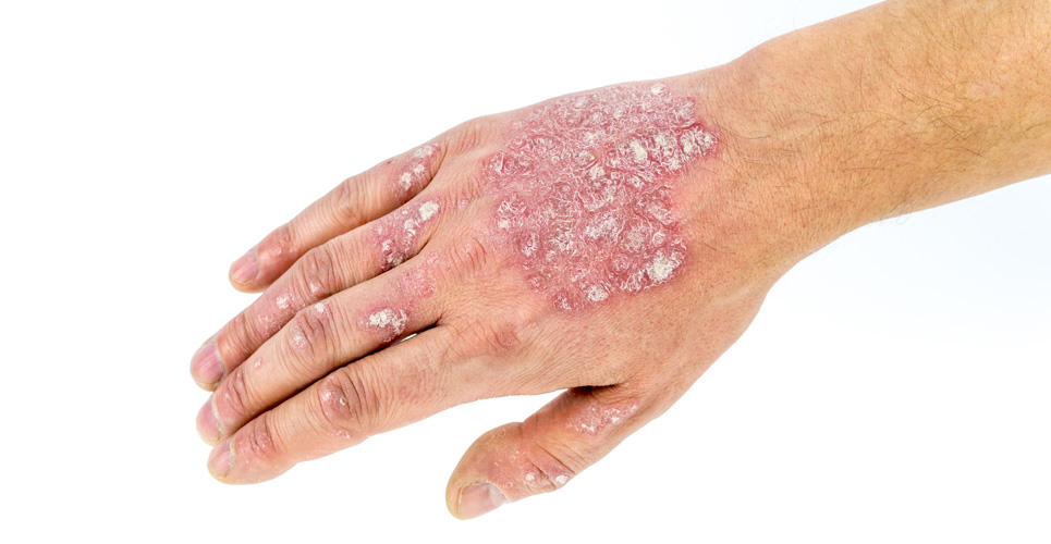
Atopic eczema (AE) is a chronic inflammatory skin disease characterised by recurrent acute and/or chronic eczematous lesions associated with pruritus. It usually starts in infancy, with an eruption of erythematous papules, oozing and crusting lesions on the face, scalp and extremities. AE may persist or occur for the first time in adolescent or adult patients, presenting with recurrent erythe‑matous rashes, scaling, lichenification and fissuring of the flexural regions. Therefore, for most patients, AE means a long-term therapy that improves both acute and chronic lesions, as well as dry skin. The aetiology of AE is complex and comprises a genetic predisposition including an impaired skin barrier function and immune responses of the host as well as environmental factors such as skin irritants, allergens and/or microbes.
Targeting skin inflammation
In acute AE lesions, there is a dense dermal infiltrate consisting of T-cells, mainly CD4+ and CD8+ cells, expressing T-helper (Th) 2 cytokines such as IL-5 and IL-13, which activate other inflammatory cells such as B-cells and eosinophils, respectively. In chronic lesions, a switch to a Th1 profile has been observed. Therefore, targeting dermal T-cells is a main approach of current therapeutic strategies.
The topical calcineurin inhibitors tacrolimus and pimecrolimus bind with high affinity to macrophilin, leading to the inhibition of the phosphatase activity of calcineurin and, consequently, reduced dephosphorylation of the nuclear factor of activated T-cells (NF-AT) cells. Dephosphorylation of NF-AT is required for its translocation to the nucleus, where it activates the transcription of various cytokine genes.(1) The clinical improvement of skin lesions upon therapy with topical calcineurin inhibitors is associated with a decrease of inflammatory cells in the dermal infiltrate as well as a reduced expression of IL-5, IL-13 and IFN-γ.(2,3) A number of clinical trials have shown that tacrolimus and pimecrolimus are effective and safe in the treatment of acute lesions, as well as in the long-term management of AE in both adults and children.(4–8)
Corticosteroids are potent anti-inflammatory drugs that target not only T-cells but also a variety of other inflammatory cells such as B-cells, monocytes/macrophages, eosinophils, dendritic cells and mast cells, as well as resident cells. Upon forming the corticosteroid/cytoplasmic glucocorticoid receptor complex and its translocation to the nucleus, the complex binds to various transcription factors, including activator protein 1 and NF-κB, resulting in the inhibition of transcriptional activity of various genes encoding proinflammatory cytokines, chemokines and adhesion molecules. Since there are concerns about side-effects in the long-term use of topical corticosteroids, they are mainly used to control acute flare-ups of AE.(9) On the other hand, long-term treatment with topical corticosteroids twice weekly prevents relapses without causing side-effects.(10)
The number of eosinophils in the dermal infiltrate was found to correlate with the degree of spongiosis in acute lesions and with epidermal hyperplasia in chronic lesions.(11) Furthermore, IL-5 expression was demonstrated in AD lesions.(12) IL-5 is known to regulate the production, differentiation, recruitment, activation and survival of eosinophils. In a placebo-controlled trial investigating the effect of an anti-IL-5 antibody in AE, 18 patients received mepolizumab. Two single doses of 750mg mepolizumab one week apart resulted in a decrease of peripheral blood eosinophil numbers, but only moderate clinical improvement of skin symptoms and pruritus was achieved as assessed by physician’s global assessment and severity scores (SCORAD, pruritus score).(13) Another study demonstrated that anti-IL-5 therapy leads to a reduction of tenascin-positive cells during the cutaneous late-phase reaction.(14) Anti-IL-5 therapy could be an approach in preventing remodelling processes in AD. However, since the pathomechanisms of AE are complex and may, in contrast to eosinophilic dermatitis, (15) not largely depend on eosinophils, blocking IL-5 alone might not be sufficient to resolve the inflammation in AE.
In 70–80% of AE patients, specific IgE antibodies against environmental allergens are detected. A humanised anti-IgE antibody, omalizumab, has been shown to be effective in the treatment of allergic diseases such as asthma, allergic rhinitis and food allergy, and may also have a potential effect in atopic dermatitis.(16,17) However, omalizumab treatment failed to change symptoms in three patients with severe AE, although one patient experienced an improvement of her bronchial asthma.(18) This report supports the concept that IgE may not be essential in the pathogenesis of AE, since approximately 20% of AE patients do not have elevated IgE levels.(2,12)
Ultraviolet (UV) phototherapy is widely used as monotherapy or in combination with topical corticosteroids in severe AE. Whereas acute lesions respond best to high- dose UVA1 or psoralen-UVA (PUVA) regimens, low-dose UVA1, 311nm UVB, UVA/UVB and broadband UVA or UVB are used in the therapy of chronic lesions.(19,20) The effect of narrow-band UVB was shown to be superior to that of broadband UVA.(21) The anti-inflammatory effects of UV light are mediated by targeting keratinocytes, epidermal dendritic cells and T-cells, resulting in a reduced expression of cytokines, which are responsible for skin inflammation and eczema formation.
Systemic immunosuppressive therapy, including ciclosporin or antimetabolites, is only used in severe, refractory AE. Because of potential side-effects, their use should be restricted to individual cases and requires close monitoring. Ciclosporin A, a potent systemic calcineurin inhibitor, improves skin symptoms in adults(22) and children(23) with AE. Upon therapy with mycophenolate mofetil, a purine biosynthesis inhibitor, a significant improvement of AE followed by a long-lasting remission has been reported. (24,25)
Skin: a barrier to the environment
Impaired skin barrier function leads to increased transepidermal water loss and dryness; in addition, the skin is more susceptible to colonisation with microbes. Daily use of moisturisers and hydration through bathing are recommended for re-establishing and preserving the barrier function. The supplementation of emollients with ceramides, the level of which is lower in AE skin, results in clinical and ultrastructural improvement of skin conditions in AE children.(26) Staphylococcus aureus is found on lesional and nonlesional skin in most AE patients. Since S aureus secretes toxins, which serve as superantigens amplifying skin inflammation, antibacterial therapy should be considered not only in patients with clinically manifest superinfections, but also in patients with moderate-to- severe AE.(27) Human manganese superoxide dismutase was shown to play a role as an autoallergen in a subset of AE patients primarily sensitised to the yeast Malassezia sympodialis.(28) Indeed, antifungal therapy was reported to be effective in AE associated with IgE-mediated hypersensitivity to yeast.(29) According to the hygiene hypothesis, the high frequency of atopic diseases is attributed to low microbial exposure in early life. Therefore, clinical trials with probiotics were initiated. Probiotics given pre- and postnatally reduced the frequency of AE in infants.(30) In children with AE, a supplementation with probiotics led to a decrease of disease severity.(31)
Conclusion
In this article we have summarised the advantages of new strategies in the treatment of AE. The treatment of AE is complex and, in addition to pharmacological therapy, it should include psychological intervention and avoidance of irritants or allergens. Future treatment options will deal with more specific drugs targeting specific cell functions – by antagonising receptors, adhesion molecules, cytokines or chemokines.
References
- Clin Exp Dermatol 2002;27:555-61.
- J Allergy Clin Immunol 2004;114:887-95.
- Allergy 2005;60:944-51.
- N Engl J Med 1997;337:816-21.
- J Am Acad Dermatol 1998;38:69-76.
- Arch Dermatol 1998;134:805-9.
- J Am Acad Dermatol 2005;52:247-53.
- Br J Dermatol 2005;152:1282-9.
- J Am Acad Dermatol 2005;53:S50-8.
- BMJ 2003;326:1367.
- Br J Dermatol 2001;145:720-9.
- J Invest Dermatol 1999;113:628-34.
- Allergy 2005;60:693-6.
- J Invest Dermatol 2004;122:1406-12.
- N Engl J Med 2003;349:2332-7.
- Clin Rev Allergy Immunol 2005;29:17-30.
- J Allergy Clin Immunol 2004;114:118-24.
- J Am Acad Dermatol 2005;53:338-40.
- Clin Exp Dermatol 2000;25:552-8.
- Clinics Dermatol 2003;21:241-8.
- Lancet 2001;357:2012-6.
- Br J Dermatol 1994;130:634-40.
- J Am Acad Dermatol 1996;34:1016-21.
- Br J Dermatol 2000;143:385-91.
- Arch Dermatol 2001;137:870-3.
- J Am Acad Dermatol 2002;47:198-208.
- Br J Dermatol 2002:147:55-61.
- J Allergy Clin Immunol 2005;115:1068-75.
- Allergy 2001;56:512-7.
- Lancet 2001;359:1076-9.
- Arch Dis Child 2005;90:881-2.