teaser
The cancer stem cells (CSC) model of tumourigenesis has the potential to revolutionise radically the way we look at malignant disease as well as the clinical management of cancer patients
Alessandro
Sgambato
MD PhD
“Giovanni XXIII” Cancer
Research Center
Institute of General
Pathology
Catholic University
Rome, Italy
The traditional view of cancer as a stochastic process in which every cell is able to initiate and maintain tumour growth has been recently challenged by the cancer stem cell model of tumourigenesis. This model postulates that tumours, like normal adult tissues, display a hierarchical structure and contain a subset of stem-like cells, the so-called cancer stem cells (CSC), being able to both self renew and give rise to a differentiated progeny which constitute the bulk of tumour.
These properties would be associated with a typical feature of stem cells: the ability to divide asymmetrically, generating two daughter cells with different properties, one identical to parent cell contributing to the maintenance of the stem cells pool and one committed to differentiate. As in other tissues, the CSC would represent only a small number of cells within a tumour and would play a pivotal role not only in the initiation and development of the primary disease but also in its recurrences and metastases. Thus, CSC are also indicated as “cancer initiating-cells” and the CSC model, if proved real, has the potentiality to impact greatly on impact our understanding of tumourigenesis as well as the clinical management of cancer patients.
CSC model: the history
It was in the first half of the 19th century that Recamier, followed by Remak, reported how cancer tissues were more similar to embryonic than adult tissues[1-2] giving support to the hypothesis that cancers arise from residues of embryonic tissue that persist into the body and do not differentiate in mature tissues with ageing.[3] It was proposed that these residual embryonic cells would possess stem-like properties, including self-renewal capacity, and would be responsible for tumour development.
The existence of such a subpopulation of tumour cells has remained theoretical until cells resembling haematopoietic stem cells were initially identified in acute myelogenous leukaemia (AML) by the observation that only a minority of leukaemic cells were able to give rise to AML with cell morphology similar to that seen in the original patients when injected in nude (immunocompromised) mice.[4,5] Afterwards, it was demonstrated that this subpopulation of cells with tumour-initiating ability could be identified and isolated by virtue of the expression of specific cell surface markers and the same method was subsequently used to isolate breast cancer cells capable of initiating the tumour in nude mice.[6] Following these initial approaches, CSC have been isolated from a variety of solid tumours as being characterised by the following biological properties: can be grown in soft agar; initiate tumours when inoculated in immunocompromised mice; are resistant to chemotherapy and radiation.
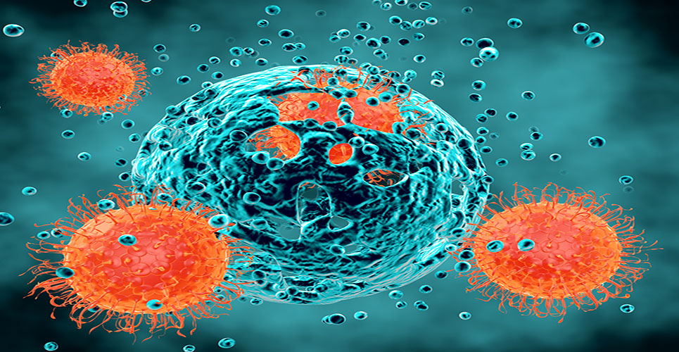
An interesting finding which recurs in most of the studies dealing with the identification of CSC in human solid tumours is the ability of the identified CSC subpopulation to recapitulate the morphological features and heterogeneity of the initial tumour when injected into nude mice and to maintain this ability after serial passages (Figure 1).
This observation is strikingly in contrast with the classical view of cancerogenesis as a stochastic event due to the accumulation of random mutations in the progeny of a single cell that initially undertakes this process. Indeed, it is highly unlikely that the same series of stochastic events occurs repetitively and exactly in the same way. Thus, the understanding of how the same cells could give rise to the same type of tumour upon each serial passage will be an important focus of cancer research in the future together with (but independently from) another fundamental open question relating to the origin of CSC. It has been hypothesised that CSC may derive from transformation of quiescent, normal long-term stem cells, recently identified in the majority of normal tissues, or could result from the de-differentiation of more mature cells.
In the first case, a normal stem cell would accumulate still unknown mutations and undergo neoplastic transformation maintaining, at least partly, its ability to give rise to various differentiated progenies thus explaining tumour cells’ heterogeneity (Figure 2). This hypothesis is in agreement with the observation that normal and cancer stem cells share similar properties and surface markers. On the other hand, it cannot be definitively excluded that CSC might derive from cells that, at some specific stages of differentiation, undergo malignant transformation thus acquiring new properties including stem-like features (Figure 2).
This hypothesis might explain the different aggressiveness of tumours which might relate to the different differentiation degree of cells undergoing the transformation event(s) as evidenced by the different tumour grading.
CSC model: CSC markers
The idea of a stem-like cell in cancers has been long suggested but the possibility to identify this putative subpopulation of tumour cells has became only recently available due to the discovery of specific cell surface markers characterising such cells. These markers are in most of the cases shared by normal stem cells of the same tissue and can be common to several histological types or more restricted (Table 1). The identification of stem cells specific markers has allowed the development of protocols, mainly based on FACS (fluorescence-activated cell sorting) or magnetic microbeads, to separate CSC from the bulk of tumour cells.
The ability to identify and isolate CSC is essential in order to be able to characterise them fully and to understand the molecular mechanisms responsible for their establishment and their maintenance. An unresolved question is the significance of CSC markers for the physiology of these cells. As shown in Table 1, mounting evidence suggests that CSCs in several human cancers are characterised by the expression of the CD133 (PROM1, prominin-1, AC133 antigen) molecule. Thus, CD133 has been considered as a bona fide CSC marker for a majority of human malignancies. However, despite the enormous interest for this molecule, its functions and ligands remain unknown and its involvement in essential properties of stem cells such as self-renewal and differentiation remains to be defined. Moreover, it is still controversial whether the identified CSC-specific markers play a direct role in stemness and tumourigenesis or they are just markers of stem-like cells with no relevant physiological functions which is an important issue in view of the possibility to develop specific anti-CSC therapies.[7]
[[hpe50.30]]
[[hpe50.31]]
CSC model and cancer therapy
The CSC model of tumourigenesis fits well with the disappointing daily experience of oncologists facing occasional complete response treatments that do not translate into cure for patients. Indeed, the model hypothesises that CSC must be eliminated to realise cures and prevent recurrences/metastases. However, CSC have been reported to be relatively resistant to standard anticancer therapies. In fact, like normal tissue stem cells, they are characterised by low rates of division and proliferation and are resistant to therapies, such as radiation and chemotherapy, which target rapidly proliferating cells. Thus, initial responses to treatment could represent therapeutic effectiveness against the bulk cancer cells while sparing CSC which would then be responsible for subsequent tumour growth both at primary and metastatic sites. This would parallel the repopulation and regeneration of tissues damaged during cancer treatment such as recovery of blood cells due to the survival of haematopoietic stem cells and re-growth of hair due to the survival of hair follicles.
[[hpe50.32]]
According to this model, a better understanding of the biology of CSC is essential to improve efficacy of anticancer therapies and several groups are focusing on this question through multiple approaches including high-throughput molecular analyses. A big problem will be to identify substantial differences between normal and cancer stem cells that might allow the development of drugs specifically targeting CSC while sparing normal stem cells with a good therapeutic index. So far, it has been demonstrated that both normal and cancer stem cells are characterised by the activation of specific molecular pathways such as WNT, Hedgehog, Bmi-1, Notch and PTEN and it has been demonstrated that targeting of CSC is possible using drugs inhibiting such pathways. However, the identification of mechanistic differences between normal and cancer stem cells will be a challenging task for the development of suitable drugs. It will be also important to identify molecular pathways responsible for therapeutic resistance of CSCs and develop specific inhibitors. In fact, besides their quiescence, CSC may also display other intrinsic resistance mechanisms due to their enhanced capacity for DNA repair and expression of efflux pumps and detoxifying enzymes. This does not mean that conventional therapies will no longer have a place in the future anticancer protocols despite the fact that CSC may be resistant to them. Indeed, it seems realistic to anticipate that a useful approach to improve current treatment of solid tumours will be the combination of a specific anti-CSC treatment (chemotherapy and/or radiation therapy) with an agent that can debulk the mass of tumour cells.[8]
Conclusions
The CSC model of tumourigenesis posits that tumours are not cellularly homogenous but contain a rare population of cells, the CSC, that display the same self-renewal, proliferative potential and cell surface markers as normal stem cells associated with the capacity to give rise to tumours and an increased resistance to therapy. Putative CSCs have now been described in the majority of human malignancies based on their ability to form tumours in immunodeficient mouse models.
However, the CSC model remains to be rigorously demonstrated in patients as well as the role of CSCs in clinical tumour growth and progression. Moreover, it remains to be definitively elucidated the relevance of CSCs in specific aspects of tumor development such as tumor angiogenesis and their relationship with the niche and the microenvironment in which they live. An important issue is the validity of assessing tumourigenic potential in immunocompromised mice for the identification of true CSCs on which some investigators still cast doubts.[9,10]
The ultimate proof of the relevance of CSCs in tumour development and in the clinical management of cancer patients will be the demonstration that specific targeting of CSCs can improve patients’ outcomes and clinical trials testing these concepts are strongly awaited. However, another important point to keep in mind is that standard response parameters might not be suitable to evaluate specific CSC-targeting therapies. Indeed, current evaluation criteria only take into consideration the effects of treatment on tumour bulk and thus might underestimate the effect of a therapy specifically targeting a rare population of cells within tumour mass. Thus, it will likely be important to re-examine the standard criteria to evaluate response therapy and other approaches, such as new CSC-specific imaging techniques, might be needed to this aim.
In conclusion, the CSC model of tumourigenesis has the potential to revolutionise radically the way we look at malignant disease as well as the clinical management of cancer patients and it is desirable to reach rapidly a definitive assessment of the role(s) that putative CSCs play in the development of human tumours and their response to therapy.
References
1. Recamier J. Recherches sur the traitement du cancer: par la compression methodique simple ou combinee, et sur l’histoire general de la meme maladie. Paris: Gabon; 1829.
2. Remak R. Deut Klin 1854;6:70-174.
3. Durante F. Arch Memor Osser Chir Pract 1874;11:217–26.
4. Lapidot T et al. Nature 1994;367:645–8.
5. Bonnet D et al. Nat Med 1997;3:730–7.
6. Al-Hajj M et al. Proc Natl Acad Sci USA 2003;100 3983–8.
7. Klonisch T et al. Trends Mol Med 2008;14:450–60.
8. Frank N et al. J Clin Invest 2010; 120:41–50.
9. Kelly P et al. Science 2007;317:337.
10. Maenhaut C et al. Carcinogenesis 2010;31:149–58.